
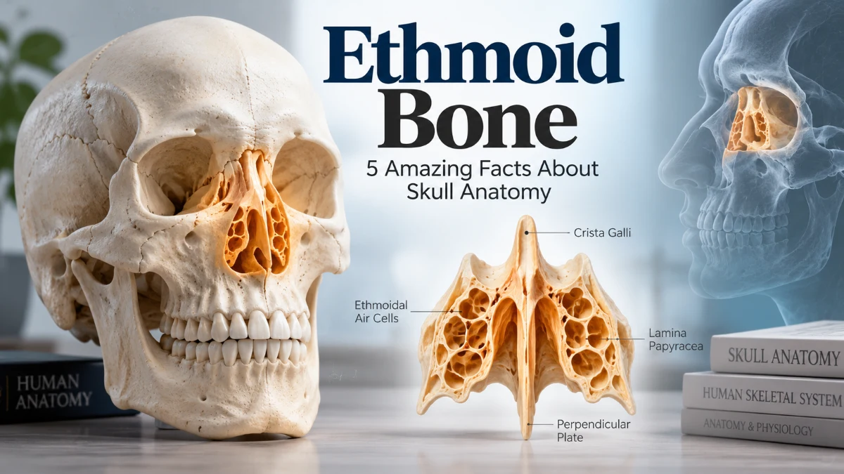
The ethmoid bone is a small, lightweight, and complex bone located deep in the central part of the skull. Despite its relatively small size, it plays an important role in the structure of the nasal cavity, nasal septum, eye sockets (orbits), and anterior cranial floor.
One of the most distinctive characteristics of the ethmoid bone is its delicate, sponge-like construction. It contains numerous small air-filled spaces known as ethmoidal air cells, which form part of the paranasal sinus system.
The ethmoid also contains the cribriform plate, a thin, perforated horizontal structure. Small bundles of olfactory receptor neuron axons pass through its tiny openings on their way from the olfactory epithelium in the nasal cavity to the olfactory bulbs. This relationship makes the ethmoid closely associated with the sense of smell.
Because of its central position and complex anatomy, the ethmoid bone is important in anatomy, medicine, radiology, otolaryngology (ENT), and surgery.
This comprehensive guide explains ethmoid bone anatomy, location, function, parts, articulations, ethmoid sinuses, clinical significance, fractures, and 10 essential facts about this fascinating skull bone.
What Is the Ethmoid Bone?
The ethmoid bone is an unpaired cranial bone located between the two orbits and at the roof of the nasal cavity.
It contributes to several important anatomical regions, including the nasal cavity, nasal septum, medial walls of the orbits, and floor of the anterior cranial fossa.
The name “ethmoid” is derived from the Greek ēthmos, meaning sieve or strainer, referring to the sieve-like appearance of the cribriform plate.
Although it is a single bone, the ethmoid contains several specialized structures with distinct anatomical functions.
Ethmoid Bone Quick Facts
| Feature | Information |
| Bone | Ethmoid bone |
| Type | Unpaired cranial bone |
| Location | Central skull, between the orbits |
| Major Parts | Cribriform plate, perpendicular plate, paired ethmoidal labyrinths |
| Important Projection | Crista galli |
| Air Spaces | Ethmoidal air cells |
| Nasal Contribution | Roof, lateral walls, and nasal septum |
| Orbital Contribution | Part of the medial wall of each orbit |
| Sensory Association | Olfaction (sense of smell) |
| Clinical Importance | Sinus disease, craniofacial trauma, CSF leaks, orbital complications |
- Read more: Ethmoid Bone
1. Where Is the Ethmoid Bone Located?
The ethmoid bone location is deep within the skull, between the right and left eye sockets and above much of the nasal cavity.
It lies near the midline and contributes to the boundary between the nasal cavity and cranial cavity.
Its central position places it in close anatomical relationship with the frontal bone, sphenoid bone, nasal structures, orbits, and anterior cranial fossa.
Unlike many other skull bones, most of the ethmoid cannot be seen easily from the external surface of an intact skull because it is positioned internally among surrounding cranial and facial structures.
Why Is the Ethmoid Bone Location Important?
The central location of the ethmoid allows it to contribute to several anatomical spaces simultaneously.
The ethmoid helps form:
- Part of the floor of the anterior cranial fossa
- Part of the roof of the nasal cavity
- Part of the nasal septum
- Portions of the lateral walls of the nasal cavity
- Part of the medial wall of each orbit
This location also explains why disease or trauma involving the ethmoid region can potentially affect the nose, sinuses, eyes, sense of smell, or intracranial structures.
2. What Are the Main Parts of the Ethmoid Bone?
Understanding the parts of the ethmoid bone makes its complex anatomy much easier to learn.
The ethmoid can broadly be divided into three major components:
- Cribriform plate
- Perpendicular plate
- Paired ethmoidal labyrinths
Each component has a distinct anatomical role.
Cribriform Plate
The cribriform plate of the ethmoid bone is a thin, horizontal, perforated structure.
It contributes to the roof of the nasal cavity and the floor of the anterior cranial fossa.
The plate contains numerous small openings known as foramina.
Small bundles of olfactory receptor neuron axons pass through these openings from the olfactory epithelium of the nasal cavity and terminate in the olfactory bulbs.
Perpendicular Plate
The perpendicular plate of the ethmoid bone projects downward from the cribriform plate.
It forms part of the superior portion of the bony nasal septum.
Inferiorly, it is related to the vomer and septal cartilage, which together contribute to the partition separating the right and left sides of the nasal cavity.
Ethmoidal Labyrinths
The paired ethmoidal labyrinths, sometimes called lateral masses, are positioned on either side of the perpendicular plate.
They contain numerous ethmoidal air cells.
The labyrinths also contribute to the medial walls of the orbits and the lateral walls of the nasal cavity.
3. What Is the Cribriform Plate?
The cribriform plate is one of the most distinctive and clinically important structures of the ethmoid bone.
The term “cribriform” means sieve-like, accurately describing its perforated appearance.
Numerous small olfactory nerve filaments pass through foramina in this plate.
These fibers collectively contribute to the olfactory nerve, or cranial nerve I (CN I), which is responsible for transmitting sensory information related to smell.
Cribriform Plate and Sense of Smell
Odor molecules stimulate specialized olfactory receptor neurons within the olfactory epithelium of the nasal cavity.
Axons from these receptor neurons travel through the small foramina of the cribriform plate and terminate in the olfactory bulbs.
Information is subsequently transmitted through additional olfactory pathways within the brain.
Because of this anatomical relationship, trauma affecting the cribriform plate and nearby olfactory fibers may potentially cause reduced or lost sense of smell.
The plate is also clinically important because it contributes to the thin boundary separating the nasal cavity from intracranial structures.
4. What Is the Crista Galli?
The crista galli is a prominent vertical projection extending upward from the cribriform plate of the ethmoid bone.
Its name means “rooster’s crest,” reflecting its distinctive appearance.
The crista galli projects into the anterior cranial fossa and serves as an important anatomical landmark.
What Is the Function of the Crista Galli?
The crista galli provides an attachment for the falx cerebri.
The falx cerebri is a fold of dura mater positioned between the right and left cerebral hemispheres.
This attachment gives the crista galli an important structural role within the cranial cavity.
The crista galli is also readily identifiable on anatomical specimens and medical imaging.
5. What Is the Function of the Ethmoid Bone?
There is no single ethmoid bone function. Instead, the bone performs several structural and sensory-supporting roles because it contributes to multiple regions of the skull.
Major Functions of the Ethmoid Bone
The ethmoid bone helps:
- Form part of the roof and lateral walls of the nasal cavity
- Form part of the bony nasal septum
- Form portions of the medial orbital walls
- Provide passage for olfactory nerve fibers
- Support structures associated with the sense of smell
- Contain the ethmoidal air cells
- Provide attachment for the falx cerebri through the crista galli
- Form the superior and middle nasal conchae
- Contribute to structures that influence airflow through the nasal cavity
Considering its relatively small size, the ethmoid contributes to a remarkable number of anatomical structures.
6. What Are the Ethmoidal Air Cells?
The ethmoidal air cells are multiple small, air-filled cavities located within the paired ethmoidal labyrinths.
Collectively, these air cells constitute the ethmoidal sinuses, which form part of the paranasal sinus system.
Unlike some other paranasal sinuses, the ethmoidal region consists of multiple individual air cells rather than a single large cavity.
Groups of Ethmoidal Air Cells
Ethmoidal air cells are generally divided into three groups:
- Anterior ethmoidal air cells
- Middle ethmoidal air cells
- Posterior ethmoidal air cells
These groups drain into different regions of the nasal cavity.
The number, size, and exact configuration of ethmoidal air cells can vary considerably among individuals.
Why Are the Ethmoid Sinuses Important?
The ethmoid sinuses are located close to the nasal cavity, orbits, and cranial cavity.
Inflammation involving the ethmoidal air cells is commonly described as ethmoid sinusitis or ethmoidal sinusitis.
Because the ethmoid sinuses are separated from the orbit by a very thin bony partition, severe infection can occasionally produce serious orbital complications.
Symptoms involving significant eye swelling, vision changes, severe headache, neurological abnormalities, or serious illness require prompt medical assessment.
7. What Are the Superior and Middle Nasal Conchae?
The ethmoid bone forms the superior nasal concha and middle nasal concha on each side of the nasal cavity.
These curved bony projections extend medially from the ethmoidal labyrinths.
An important anatomical distinction is that the inferior nasal concha is a separate facial bone and is not part of the ethmoid bone.
What Is the Function of the Nasal Conchae?
The nasal conchae increase the internal surface area of the nasal cavity and influence the movement of inhaled air.
Together with the surrounding nasal mucosa, they contribute to warming, humidifying, and filtering inspired air.
The curved conchae also create passageways known as nasal meatuses.
These spaces are important because several paranasal sinus drainage pathways communicate with different areas of the nasal cavity.
8. How Does the Ethmoid Bone Form Part of the Orbit?
A very thin portion of each ethmoidal labyrinth contributes to the medial wall of the orbit.
This thin bony structure is commonly known as the lamina papyracea, or orbital plate of the ethmoid.
Its name reflects its remarkably thin, paper-like appearance.
Why Is the Lamina Papyracea Clinically Important?
The lamina papyracea forms an important boundary between the ethmoidal air cells and the orbit.
Because this partition is extremely thin, infection or disease involving the ethmoid sinuses can potentially spread toward orbital tissues.
This anatomical relationship is particularly important when healthcare professionals evaluate complications of severe sinus infections.
The thinness of this area is also relevant in facial trauma and during certain endoscopic sinus and orbital procedures.
9. Which Bones Articulate With the Ethmoid Bone?
The ethmoid bone articulations demonstrate how centrally positioned this bone is within the skull.
The ethmoid articulates with numerous cranial and facial bones.
These include the:
- Frontal bone
- Sphenoid bone
- Vomer
- Two nasal bones
- Two maxillae
- Two lacrimal bones
- Two palatine bones
- Two inferior nasal conchae
These articulations integrate the ethmoid with the cranial base, nasal cavity, orbits, and facial skeleton.
Ethmoid Bone and Vomer Relationship
Students sometimes confuse the perpendicular plate of the ethmoid with the vomer because both contribute to the bony nasal septum.
The perpendicular plate of the ethmoid forms much of the superior portion of the bony septum.
The vomer contributes substantially to its posteroinferior portion.
The septal cartilage forms a major anterior component.
Remembering these relationships can make the anatomy of the nasal septum easier to understand.
10. Why Is the Ethmoid Bone Clinically Important?
The ethmoid bone anatomy is clinically important because several delicate and vital structures lie close together in this region.
Conditions involving the ethmoid may potentially affect:
- The sense of smell
- Paranasal sinus drainage
- The nasal cavity
- The orbit and surrounding tissues
- The boundary between the nasal and cranial cavities
This explains why the ethmoid is particularly relevant in ENT medicine, radiology, ophthalmology, trauma assessment, and skull-base surgery.
Ethmoid Bone Fracture
An ethmoid bone fracture can occur as part of significant craniofacial or skull-base trauma.
The effects depend on the location and severity of the injury.
Damage involving the cribriform plate can be particularly concerning because this thin region contributes to the separation between the nasal cavity and intracranial space.
Potential findings after significant injury may include changes in the sense of smell or leakage of cerebrospinal fluid.
Suspected skull-base trauma requires prompt professional medical assessment.
Cerebrospinal Fluid Leak
Damage involving the cribriform plate may sometimes create an abnormal communication between the intracranial and nasal spaces.
This can result in cerebrospinal fluid (CSF) rhinorrhea, in which CSF drains through the nose.
Clear or watery nasal drainage after significant head trauma should therefore not automatically be assumed to be ordinary nasal mucus.
A suspected CSF leak following trauma requires urgent medical evaluation because proper diagnosis and management are important.
Loss of Smell
Because olfactory nerve fibers pass through the cribriform plate, trauma in this region may damage these delicate fibers.
Possible consequences include hyposmia, meaning a reduced sense of smell, or anosmia, meaning loss of smell.
However, changes in smell can have many different causes. Loss of smell alone does not establish that an ethmoid fracture is present.
Ethmoid Bone vs Sphenoid Bone
The ethmoid and sphenoid are both centrally positioned cranial bones, but their anatomy and major functions differ substantially.
| Feature | Ethmoid Bone | Sphenoid Bone |
| General Location | Between the orbits and around the upper nasal cavity | Central region of the cranial base |
| Notable Feature | Cribriform plate | Sella turcica |
| Air Spaces | Ethmoidal air cells | Sphenoidal sinuses |
| Nasal Contribution | Extensive | More limited |
| Orbital Contribution | Primarily medial orbital wall | Contributes to several orbital regions |
| Olfactory Relationship | Olfactory fibers pass through cribriform plate | Not the principal passage for olfactory fibers |
Both bones nevertheless contribute to the complex architecture of the cranial base and articulate with one another.
Ethmoid Bone vs Vomer
Another useful anatomical comparison is the ethmoid bone vs vomer.
Both participate in formation of the bony nasal septum, but they are distinctly different bones.
The perpendicular plate of the ethmoid contributes mainly to the superior portion of the bony nasal septum.
The vomer contributes substantially to the posteroinferior portion.
The ethmoid is considerably more complex and also includes the cribriform plate, crista galli, ethmoidal labyrinths, ethmoidal air cells, and superior and middle nasal conchae.
The vomer, by comparison, is a separate facial bone primarily associated with formation of the nasal septum.
How to Remember Ethmoid Bone Anatomy
The easiest way to remember ethmoid bone anatomy is to visualize its major structures according to direction.
Think of the cribriform plate as the horizontal, perforated component.
The crista galli projects upward from the midline of the cribriform plate.
The perpendicular plate projects downward and contributes to the nasal septum.
The ethmoidal labyrinths are positioned on either side and contain the ethmoidal air cells.
Also remember the simple relationship:
Up = crista galli
Across = cribriform plate
Down = perpendicular plate
Sides = ethmoidal labyrinths
Finally, remember that the ethmoid links four important anatomical areas: the nose, sinuses, orbits, and anterior cranial cavity.
Clinical Significance of Ethmoid Bone Anatomy
The ethmoid region can be evaluated using medical imaging, particularly computed tomography (CT), when clinically appropriate.
CT imaging can provide detailed information about the ethmoidal air cells, surrounding bony structures, nasal cavity, orbit, and skull base.
Healthcare professionals may evaluate this region in cases involving sinus disease, facial trauma, orbital complications, suspected skull-base abnormalities, or surgical planning.
Detailed knowledge of ethmoid anatomy is particularly important during certain endoscopic sinus and skull-base procedures.
Surgeons working in this region must understand the close relationships among the nasal cavity, ethmoid sinuses, orbit, cribriform plate, and intracranial structures.
Individual anatomical variation also makes careful preoperative assessment important for appropriate procedures.
Frequently Asked Questions
What is the ethmoid bone?
The ethmoid bone is an unpaired cranial bone located centrally between the eye sockets and above much of the nasal cavity. It contributes to the nasal cavity, nasal septum, medial orbital walls, and floor of the anterior cranial fossa.
Where is the ethmoid bone located?
The ethmoid bone is located deep within the skull between the two orbits and superior to much of the nasal cavity. It lies near the anterior cranial fossa.
What are the three main parts of the ethmoid bone?
The three major components are the cribriform plate, perpendicular plate, and paired ethmoidal labyrinths.
What is the main function of the ethmoid bone?
The ethmoid has several functions rather than one single function. It helps form the nasal cavity, nasal septum, and medial orbital walls, contains ethmoidal air cells, and provides passage for olfactory nerve fibers.
What passes through the cribriform plate of the ethmoid bone?
Small bundles of olfactory receptor neuron axons pass through foramina in the cribriform plate and terminate in the olfactory bulbs. These fibers collectively contribute to the olfactory nerve, or cranial nerve I.
Is the ethmoid bone part of the nasal septum?
The perpendicular plate of the ethmoid bone contributes to the superior portion of the bony nasal septum.
Does the ethmoid bone contain sinuses?
The paired ethmoidal labyrinths contain numerous ethmoidal air cells, which collectively constitute the ethmoidal sinuses and form part of the paranasal sinus system.
What is the crista galli?
The crista galli is an upward-projecting portion of the ethmoid bone. It provides an attachment for the falx cerebri, a fold of dura mater positioned between the cerebral hemispheres.
Can an ethmoid bone fracture affect the sense of smell?
Yes, potentially. Trauma involving the cribriform plate and nearby olfactory nerve fibers may cause reduced or lost sense of smell. The effects depend on the nature and severity of the injury.
Why is the ethmoid bone clinically important?
The ethmoid bone is clinically important because it lies close to the nasal cavity, paranasal sinuses, orbits, olfactory structures, and cranial cavity. Consequently, trauma, infection, and surgical procedures in this region require careful consideration of surrounding structures.