
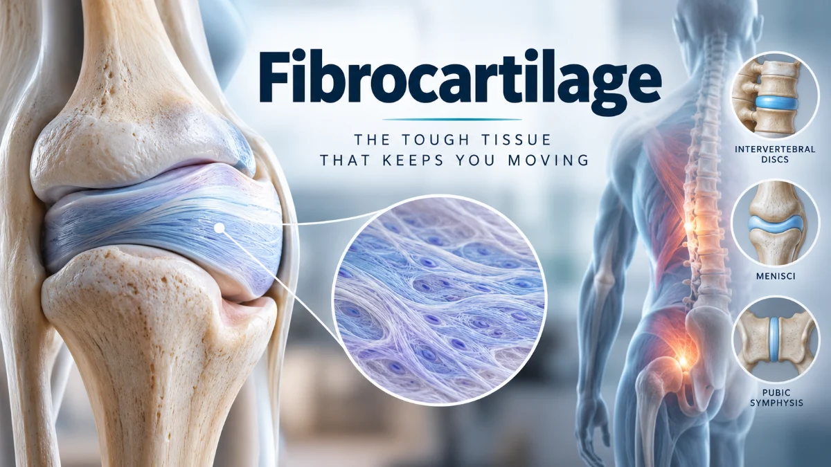
Fibrocartilage is a tough, specialized type of cartilage that provides structural support, absorbs shock, and helps joints withstand mechanical stress. It contains thick bundles of collagen fibers and is found in important structures such as the knee menisci, intervertebral discs, pubic symphysis, and certain joint discs.
Unlike hyaline cartilage, which forms smooth surfaces in many joints, fibrocartilage is adapted to resist substantial tensile and compressive forces. Its collagen-rich structure makes it particularly important in areas subjected to repeated pressure, stretching, and movement.
This connective tissue contributes to everyday activities such as walking, running, bending, and lifting. However, injuries and degenerative changes can affect its function, and its ability to repair itself is limited.
This comprehensive guide explains fibrocartilage anatomy, structure, functions, locations, histology, healing potential, common injuries, and treatment options. It also compares the three major types of cartilage and answers 10 frequently asked questions.
Medical Disclaimer: This article is for general educational purposes only and does not replace professional medical advice, diagnosis, or treatment. Seek medical evaluation for persistent pain, significant swelling, restricted movement, or a suspected joint injury.
Quick Facts About Fibrocartilage
| Feature | Description |
| Tissue type | Specialized connective tissue |
| Main collagen | Predominantly type I collagen |
| Additional collagen | Type II collagen, in varying amounts |
| Main cells | Chondrocytes and fibrochondrocytes |
| Primary functions | Strength, shock absorption, and stability |
| Common locations | Knee menisci, intervertebral discs, pubic symphysis |
| Perichondrium | Absent |
| Blood supply | Generally limited; varies by location |
| Healing capacity | Limited and dependent on the injury |
Fibrocartilage combines characteristics of dense connective tissue and cartilage, allowing it to resist tension and compression.
Kenhub
1. What Is Fibrocartilage?
Fibrocartilage is one of the three major types of cartilage in the human body. The other two are hyaline cartilage and elastic cartilage.
It is composed of specialized cartilage cells surrounded by a dense extracellular matrix containing collagen fibers, proteoglycans, and water.
Its structure provides mechanical strength while allowing a limited degree of flexibility.
Fibrocartilage Definition in Anatomy
In anatomy, fibrocartilage is a type of cartilage characterized by abundant type I collagen fibers and chondrocytes located within a dense extracellular matrix.
It is particularly well suited for anatomical regions exposed to substantial mechanical loading.
Key Characteristics
Fibrocartilage has several distinctive characteristics:
- Thick type I collagen bundles provide tensile strength.
- Type II collagen is present in varying amounts.
- Chondrocytes occupy small spaces called lacunae.
- The tissue resists compression, tension, and shear forces.
- It lacks a perichondrium.
- Its natural healing capacity is generally limited.
These structural features distinguish it from other cartilage types.
- Read more: Fibrocartilage
2. Fibrocartilage Structure and Composition
The microscopic structure of fibrocartilage explains its ability to tolerate considerable mechanical stress.
It consists primarily of collagen fibers, specialized cells, and extracellular matrix.
Collagen Fibers
Type I collagen is particularly abundant in fibrocartilage.
It forms thick bundles that provide resistance to stretching and pulling forces.
Type II collagen may also be present, although its proportion varies depending on the anatomical location.
The arrangement of collagen fibers reflects the mechanical demands placed on the tissue.
For example, collagen organization within knee menisci helps distribute forces generated during weight-bearing activities.
Chondrocytes and Fibrochondrocytes
Chondrocytes are specialized cells responsible for maintaining cartilage matrix.
They are located within microscopic cavities called lacunae.
These cells often appear in rows between collagen bundles.
Fibrocartilage also contains specialized cells called fibrochondrocytes, which display characteristics associated with both fibroblasts and chondrocytes.
These cells contribute to the maintenance of the surrounding matrix.
Extracellular Matrix
The extracellular matrix contains:
- Collagen fibers
- Proteoglycans
- Glycosaminoglycans
- Water
- Other structural components
Proteoglycans help retain water, contributing to the tissue’s resistance to compression.
The relative proportions of these components differ among anatomical locations.
Does Fibrocartilage Have a Perichondrium?
No. Fibrocartilage lacks a perichondrium, the connective tissue covering found around many other cartilage structures.
Its limited vascularity and specialized cellular environment contribute to its restricted repair capacity.
However, the blood supply of individual fibrocartilaginous structures can vary.
3. Where Is Fibrocartilage Found in the Body?
Fibrocartilage is primarily found in structures that require strength, stability, and resistance to mechanical loading.
Its location reflects the need to distribute pressure and withstand forces generated during movement.
Main Locations of Fibrocartilage
| Anatomical location | Primary function |
| Intervertebral discs | Distribute forces between vertebrae |
| Knee menisci | Distribute weight and absorb shock |
| Pubic symphysis | Support pelvic stability |
| Temporomandibular joint disc | Support jaw movement and load distribution |
| Wrist triangular fibrocartilage complex | Support wrist stability |
| Hip and shoulder labra | Improve joint stability |
Intervertebral Discs
Intervertebral discs are located between most adjacent vertebral bodies in the spine.
Each disc consists of a tough outer annulus fibrosus and a softer, hydrated inner nucleus pulposus.
The annulus contains layered fibrocartilaginous tissue that helps resist tension and distribute mechanical loads.
Together, these structures permit controlled spinal movement and help distribute pressure during activities such as walking and lifting.
Knee Menisci
The knee contains two menisci:
- Medial meniscus
- Lateral meniscus
These crescent-shaped fibrocartilaginous structures lie between the femur and tibia.
They help distribute body weight, improve joint stability, and reduce concentrated pressure on the articular cartilage.
Damage to a meniscus can affect normal knee movement and load distribution.
Pubic Symphysis
The pubic symphysis connects the left and right pubic bones at the front of the pelvis.
A fibrocartilaginous disc separates the bones and permits a small amount of movement.
This structure helps maintain pelvic stability while transmitting mechanical forces between the two sides of the pelvis.
Temporomandibular Joint
The temporomandibular joint connects the mandible to the skull.
Its articular disc contains specialized fibrocartilaginous tissue that helps coordinate jaw movement and distribute forces during chewing and speaking.
Other Locations
Fibrocartilaginous tissue is also present in the triangular fibrocartilage complex of the wrist, the hip and shoulder labra, and certain tendon attachment sites.
Its exact composition varies according to the mechanical demands of each structure.
4. What Is the Function of Fibrocartilage?
The main function of fibrocartilage is to provide strength and structural support in areas exposed to considerable mechanical stress.
However, it performs several important functions.
Shock Absorption
Fibrocartilaginous structures help absorb and distribute mechanical forces.
For example, the knee menisci spread loads across the joint, reducing concentrated pressure on the underlying articular surfaces.
Intervertebral discs perform a related function by distributing pressure between vertebrae.
Resistance to Compression
Compression occurs when forces push a structure together.
Fibrocartilage resists compressive loading through its collagen organization and hydrated extracellular matrix.
This property is particularly important in weight-bearing structures.
Tensile Strength
Tensile stress develops when tissues are stretched or pulled.
Thick bundles of type I collagen provide resistance to these forces and help maintain structural integrity.
Joint Stability
Certain fibrocartilaginous structures improve joint stability.
The hip and shoulder labra deepen their respective joint sockets, while the knee menisci contribute to joint congruence and load distribution.
Structural Support
Fibrocartilage reinforces anatomical structures exposed to repetitive mechanical loading.
Its mechanical properties help support the spine, pelvis, and several important joints.
5. Fibrocartilage vs. Hyaline Cartilage vs. Elastic Cartilage
The human body contains three major types of cartilage.
Although they share some cellular characteristics, their composition, structure, and mechanical properties differ.
Cartilage Comparison Table
| Feature | Fibrocartilage | Hyaline cartilage | Elastic cartilage |
| Main fibers | Type I collagen, with variable type II | Type II collagen | Type II collagen and elastic fibers |
| Appearance | Dense and fibrous | Smooth, glass-like matrix | Elastic fiber network |
| Main function | Strength and shock absorption | Smooth joint movement and support | Flexible support |
| Flexibility | Relatively limited | Moderate | High |
| Common locations | Menisci, discs, pubic symphysis | Joint surfaces, nose, trachea | External ear, epiglottis |
| Perichondrium | Absent | Usually present, except at articular surfaces | Usually present |
The three cartilage types are adapted to different mechanical and anatomical requirements.
Kenhub
Is Fibrocartilage the Strongest Type of Cartilage?
Fibrocartilage is generally considered the toughest of the three major cartilage types because of its dense collagen network and high tensile strength.
However, mechanical strength depends on the type of force involved.
Hyaline cartilage provides smooth, low-friction joint surfaces, while elastic cartilage offers flexible structural support.
Each type performs a specialized function.
6. Fibrocartilage Histology: How to Identify It Under a Microscope
Histology is the microscopic study of biological tissues.
Fibrocartilage can be identified by examining its collagen fibers, cartilage cells, and extracellular matrix.
Microscopic Features
A typical fibrocartilage specimen contains thick collagen bundles arranged in parallel or layered patterns.
Chondrocytes appear within lacunae and are frequently arranged in rows between the collagen fibers.
The tissue generally appears more fibrous than hyaline cartilage.
Hematoxylin and Eosin Staining
Hematoxylin and eosin (H&E) is a commonly used staining method in histology.
With this technique:
- Collagen-rich regions generally appear pink.
- Cell nuclei appear blue or purple.
- Chondrocytes are visible within lacunae.
- Thick collagen bundles are relatively conspicuous.
The exact appearance depends on the specimen, tissue orientation, and staining method.
Fibrocartilage vs. Hyaline Cartilage Histology
Hyaline cartilage has a comparatively smooth, glass-like matrix under routine light microscopy.
Fibrocartilage displays visible collagen bundles with cartilage cells distributed between them.
Another characteristic feature is the absence of a perichondrium.
However, this feature is not unique to fibrocartilage because articular hyaline cartilage also lacks a perichondrium.
7. Common Fibrocartilage Injuries and Disorders
Although fibrocartilage is strong, it can become damaged through trauma, repetitive mechanical loading, or degenerative changes.
Symptoms depend on the affected anatomical structure and injury severity.
Meniscus Tears
A meniscus tear is a common knee injury involving fibrocartilaginous tissue.
It may occur during sudden twisting movements or develop gradually through degenerative changes.
Common symptoms include:
- Knee pain
- Swelling and stiffness
- Clicking or catching
- Difficulty straightening the knee
- A sensation of instability
Not every meniscal tear causes significant symptoms or requires surgery.
Treatment depends on the tear’s location, characteristics, symptoms, and the patient’s circumstances.
Intervertebral Disc Injuries
Intervertebral discs may develop structural changes associated with aging, repetitive loading, or injury.
Disc degeneration can involve changes in hydration and the structure of the annulus fibrosus.
A herniated disc occurs when disc material becomes displaced beyond its normal boundaries.
Some disc abnormalities can irritate nearby nerves and cause pain, numbness, or weakness.
However, imaging abnormalities do not always cause symptoms, so clinical assessment remains important.
Wrist Fibrocartilage Injuries
The triangular fibrocartilage complex (TFCC) supports wrist stability and load distribution.
Injuries can occur after falls, twisting movements, or repetitive mechanical loading.
Symptoms may include pain on the little-finger side of the wrist, clicking, and reduced grip strength.
Labral Injuries
The hip and shoulder contain fibrocartilaginous labra that contribute to joint stability.
Labral injuries may cause pain, catching, or instability.
However, these symptoms are not specific to labral damage, and a healthcare professional must evaluate the underlying cause.
8. Can Fibrocartilage Heal Naturally?
Fibrocartilage has a limited natural healing capacity because much of the tissue lacks a direct internal blood supply.
Blood delivers oxygen, nutrients, and cells involved in tissue repair.
Consequently, injuries in poorly vascularized regions may heal slowly or fail to regenerate completely.
Blood Supply and Healing
The knee meniscus illustrates how blood supply influences healing.
Its outer region receives blood from surrounding tissues, while the inner region has considerably less vascularity.
Tears in better-vascularized regions may have greater healing potential than tears in poorly vascularized areas.
However, healing also depends on the tear pattern, stability, tissue quality, and other factors.
Can Small Meniscus Tears Heal Without Surgery?
Some small, stable meniscal tears can heal or become asymptomatic with appropriate nonsurgical management.
However, improvement in symptoms does not necessarily mean that the original tissue has fully regenerated.
Treatment decisions should consider the patient’s symptoms, activity requirements, and injury characteristics.
How Long Does Fibrocartilage Take to Heal?
There is no universal healing timeline.
Recovery depends on the affected anatomical structure, injury severity, treatment method, and individual circumstances.
Some patients regain comfortable movement through rehabilitation, while others require surgical treatment or prolonged management.
The healing characteristics of the knee meniscus should not be assumed to apply to every fibrocartilaginous structure.
9. Fibrocartilage Injury Diagnosis and Treatment
Diagnosis usually begins with a medical history and physical examination.
A healthcare professional may assess pain, swelling, tenderness, movement, and joint stability.
Treatment depends on the specific injury rather than fibrocartilage damage alone.
Physical Examination
For suspected meniscal injuries, clinicians may use specialized knee examination techniques.
These tests help identify possible damage and determine whether additional imaging is necessary.
Imaging Tests
Magnetic resonance imaging (MRI) provides detailed images of soft tissues, including knee menisci and intervertebral discs.
X-rays are useful for examining bones and certain joint abnormalities, but they do not directly display soft cartilage as clearly as MRI.
The American Academy of Orthopaedic Surgeons recommends MRI as the preferred imaging modality for diagnosing acute isolated meniscal tears because of its diagnostic accuracy. Its guideline specifically addresses acute isolated injuries rather than all types of meniscal damage.
Nonsurgical Treatment
Depending on the diagnosis, nonsurgical treatment may include:
- Activity modification
- Physiotherapy
- Strengthening exercises
- Appropriate pain management
- Temporary bracing or other support when recommended
The goal is to improve function, manage symptoms, and support safe recovery.
Some patients recover without surgery.
Surgical Treatment
Surgery may be considered for selected injuries, particularly certain repairable tears that cause substantial symptoms or mechanical locking.
Meniscal procedures may include surgical repair or selective removal of damaged tissue.
When surgery is indicated for an acute meniscal tear, preserving as much functional meniscal tissue as possible is an important treatment objective.
When Should You See a Doctor?
Seek medical evaluation if you experience persistent joint pain, significant swelling, locking, or difficulty bearing weight after an injury.
Seek emergency assessment if back pain is accompanied by new bladder or bowel dysfunction, numbness around the groin or saddle region, or rapidly worsening leg weakness.
These symptoms may indicate serious neurological involvement requiring urgent evaluation.
10. How to Maintain Healthy Fibrocartilage
No lifestyle strategy can prevent every cartilage injury. However, several habits can support overall musculoskeletal health and reduce avoidable mechanical stress.
Exercise Regularly
Regular physical activity supports muscle strength, coordination, and joint mobility.
Depending on individual circumstances, walking, swimming, cycling, and strength training may be suitable.
Exercise intensity should increase gradually.
Strengthen Supporting Muscles
Strong muscles help stabilize joints and manage mechanical forces.
For example, strengthening the quadriceps, hamstrings, and hip muscles can support knee function.
People recovering from injuries may benefit from exercises prescribed by a physiotherapist.
Maintain an Appropriate Body Weight
Body weight influences mechanical loading on weight-bearing joints.
Weight management, when medically appropriate, may reduce some of this loading.
However, joint health also depends on genetics, previous injuries, muscle strength, and other factors.
Increase Activity Gradually
Sudden increases in exercise intensity can expose tissues to unfamiliar mechanical loads.
Progressive training, proper techniques, and adequate recovery may help reduce avoidable injuries.
Follow a Balanced Diet
Adequate protein, vitamins, minerals, and energy support general tissue maintenance.
However, no specific food or supplement has been established as a reliable method for completely regenerating damaged human fibrocartilage.
Products promising guaranteed cartilage regrowth should be approached cautiously.
Follow Rehabilitation Instructions
After an injury, follow the recovery plan recommended by your healthcare professional.
Returning to demanding activities too early may increase the risk of additional damage.
Frequently Asked Questions About Fibrocartilage
What is fibrocartilage in simple terms?
Fibrocartilage is a tough type of cartilage containing thick collagen fibers. It provides strength, absorbs shock, and supports structures such as the knee menisci and intervertebral discs.
Where is fibrocartilage found in the human body?
It is commonly found in the knee menisci, intervertebral discs, pubic symphysis, temporomandibular joint disc, and specialized structures within the wrist, hip, and shoulder.
What is the main function of fibrocartilage?
Its main functions are providing structural strength, resisting mechanical stress, distributing pressure, absorbing shock, and supporting joint stability.c
Which type of collagen is found in fibrocartilage?
Type I collagen is particularly abundant in fibrocartilage. Type II collagen may also be present in varying amounts depending on the anatomical location.
Does fibrocartilage have a blood supply?
Fibrocartilage generally lacks an internal blood supply, although vascularity varies by structure. The outer portion of the knee meniscus receives blood from surrounding tissues.
Why does fibrocartilage heal slowly?
Its limited blood supply restricts the delivery of blood-derived cells and other components involved in repair. Healing also depends on injury location, size, stability, and tissue condition.
What is the difference between fibrocartilage and hyaline cartilage?
Fibrocartilage contains prominent type I collagen bundles and provides tensile strength and shock absorption. Hyaline cartilage contains predominantly type II collagen and provides smooth joint surfaces and structural support.
Is fibrocartilage found in the knee?
Yes. The medial and lateral menisci are fibrocartilaginous structures that help distribute weight, absorb impact, and improve knee stability.
Can damaged fibrocartilage regenerate?
Natural regeneration is limited. Some injuries in better-vascularized regions may heal, while others require rehabilitation or surgery. Complete restoration of the original tissue is not always possible.
What happens when fibrocartilage is damaged?
Damage may cause pain, swelling, restricted movement, or joint instability, depending on the affected structure. Some abnormalities cause few symptoms, while others require medical assessment and treatment.